

Facelift
General information
Anesthesia
routinely general (general anesthesia)
Duration
5-6 h
Stay in clinic
36 h (surgery on Monday morning, discharge on Tuesday late afternoon)
Follow-up examinations
- The day after the procedure – removal of drains from the wound, local condition check, application of a supportive garment.
- After one week (mandatory) – local condition check, partial removal of sutures.
- After 2 weeks (mandatory) – local condition check, removal of remaining sutures.
- After a longer period from the procedure (preferably 1 year after) – final assessment of the surgery result.
- Additionally, at any time when needed (optional).
Recovery period
- Early recovery period 2-3 weeks (better to reserve 3 weeks).
- Sleeping on your back with your head slightly elevated for 2 weeks.
- Washing hair after 3 days, once a day (with a gentle baby shampoo).
- Avoiding hard and hot foods for several days.
- Return to intellectual work after 2-3 weeks (after this time, bruising and significant swelling usually disappear);
- Return to physical exertion and light sport after 1 month.
- Protect facial skin from sun radiation and severe frost for 1 year.
Medication after the procedure
Over-the-counter painkillers.
Wearing a special compression garment on the face for 1 week non-stop (with a break for washing hair), for another week only at night.
Inevitable consequences of the procedure (always occur)
- pain/discomfort (mainly in the form of pulling, feeling of tension);
- swelling;
- bruising;
- sensory disturbances in the area of the surgical wound;
- scar;
- a facelift is an operation that does not yield a perfect result – improvement can be achieved, but there are always minor imperfections of the procedure after surgery.
Surgical complications (may occur)
- hematoma (accumulation of blood requiring surgery or needle aspiration);
- infection (requires antibiotics);
- prolonged wound healing (requires additional check-ups and dressings);
- hypertrophic scar/keloid (rare);
- necrosis of the skin-fat flap (very rare);
- paresis or paralysis of one of the branches of the facial nerve (very rare);
- prolonged postoperative pain;
- the face before the procedure is always asymmetrical (it is also asymmetrical after the procedure);
- dissatisfaction with the result of the procedure;
- overcorrection (after some time, overly tense tissues relax spontaneously);
- undercorrection (usually requires another procedure);
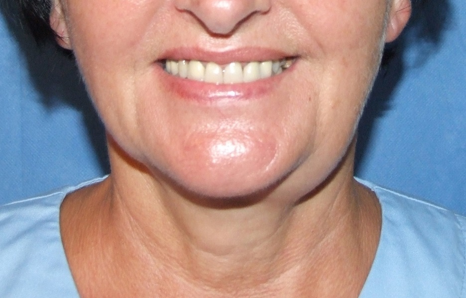
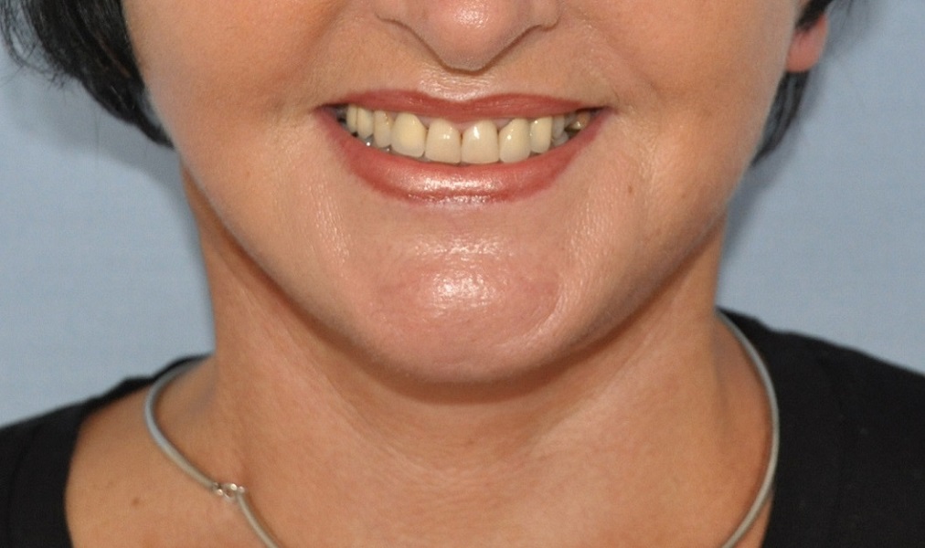
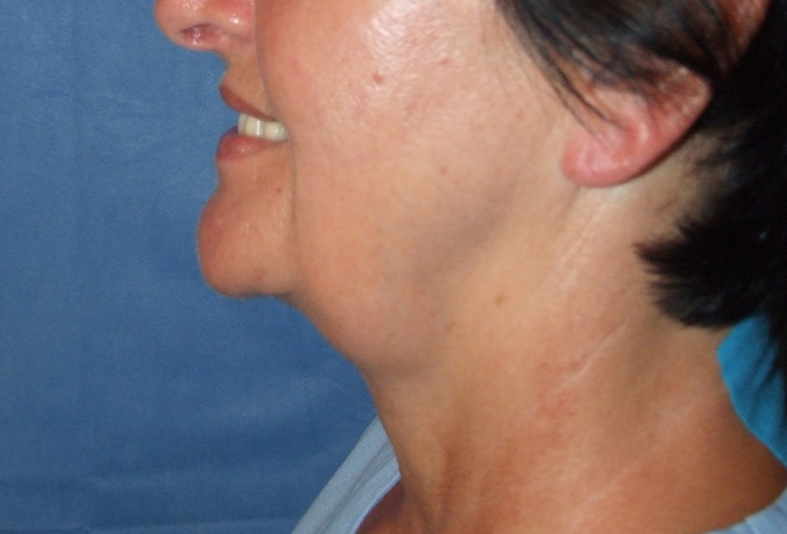
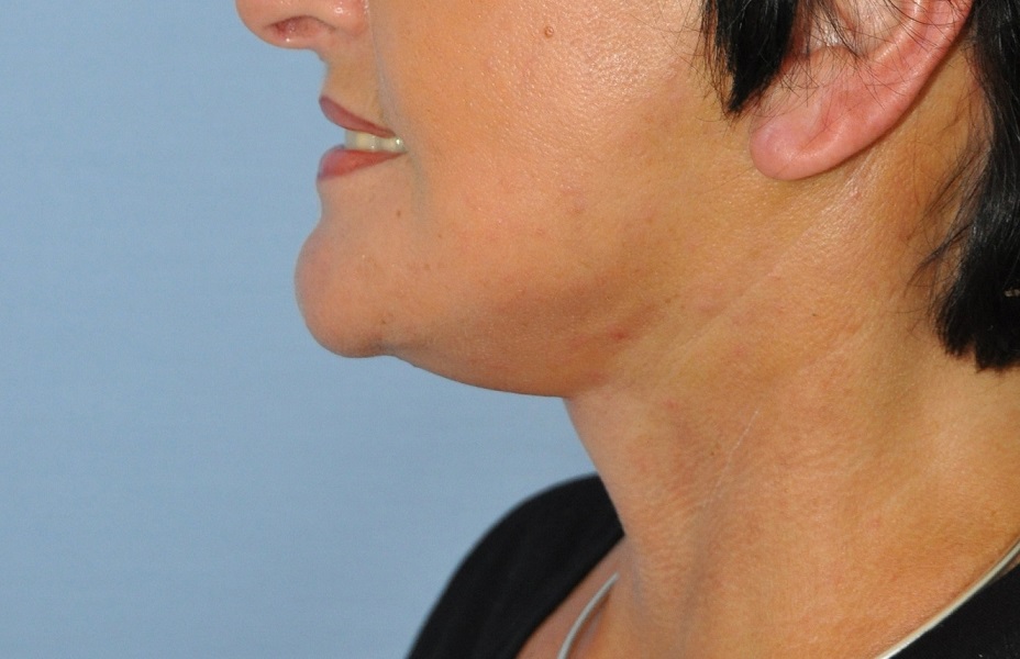
Photo gallery
Detailed information
Introduction
The basis of all plastic surgery procedures is knowledge of normal and functional anatomy, as well as aesthetic norms related to the human body. Aesthetic norms are created based on the feeling and perception of human bodily beauty. Beauty is a positive aesthetic quality of being, resulting from the preservation of proportion, harmony, moderation, and utility (functionality). In Western culture, recognized patterns of bodily beauty in sculpture include: „Venus de Milo” and Michelangelo's „David,” and in painting – Botticelli's „The Birth of Venus.” Beauty is determined by youth, symmetry, and gender specificity: a woman has features specific to femininity, and a man to masculinity. Ugliness, as the opposite of beauty, is attributed to phenomena and objects that are not beautiful according to the canons accepted in a given place and time. Ugliness in art is represented, for example, by the Hellenistic sculpture „Old Drunkard” and Albrecht Dürer's painting „Old Prostitute.” A common feature of ugliness in the depicted figures is old age.
Appearance is of great importance in human life. How we perceive another person depends 80% on their appearance. People considered attractive, „beautiful,” have a life advantage over others. Plastic surgeons restore external features to people that nature gave them, and which they lost as a result of injury, illness, or the passage of time. The measure of benefit gained by patients is not only an objectively better appearance but also an improvement in their mental state.
Aging, along with the subsequent loss of function, is reflected in the anatomical state of tissues. Plastic surgery has tools that allow influencing anatomy to remove external signs of aging.
Normal and topographical anatomy and the selection of treatment for a given patient
The human body is built in layers. The foundation is the bone framework – it determines the shape of the face as a whole. The mandibular skeleton en face can fit into a contour similar to a triangle or a square. The former is characteristic of a female face, the latter – of a male face. Regardless of gender, the contour of a young face en face fits into a gentle oval shape. Generally speaking, a facelift is aimed at restoring the oval of the face. In profile, the jawline and neck line form the mentocervical angle. It can be deep and close to a right angle, or shallow and obtuse. A deep and sharp angle is desirable from the perspective of aesthetic norms. Soft tissues are suspended on the bone framework. Going successively from deeper to more superficial, these are: the skeletal muscle layer, deep fascia, then subcutaneous tissue with superficial fascia, and skin. The superficial fascial layer on the head and neck takes the form of SMAS (Superficial Musculo-Aponeurotic System). In the inferolateral part of the face, the visible skeletal muscle is the masseter muscle (its prominence can be influenced using botulinum toxin, achieving an improvement in the contour of the entire face). In the neck, a deep and superficial layer of skeletal neck muscles can be distinguished (platysma – the broad muscle of the neck is part of the SMAS, not an element of the superficial layer of skeletal neck muscles). A distinct and always visible skeletal muscle of the superficial layer is the sternocleidomastoid muscle. The anterior belly of the digastric muscle (another skeletal neck muscle of practical importance in a facelift) can become too prominent as a result of aging processes, and a procedure on it can be an element of restoring the neck contour.
In the lateral part of the chin, behind the body of the mandible, there is the submandibular salivary gland, covered only by SMAS and skin. With aging processes, it can lower its position and significantly affect the contour of the chin. Reduction of its mass can be an element of improving the contour of the jaw and neck. The parotid gland also plays a role in face and neck lifts, although its anatomy is not modified during surgery.
The SMAS layer is not homogeneous throughout the head and neck. In some places, it is fibro-fatty, in others, musculo-aponeurotic. In some areas, SMAS is strongly attached to the bone framework, in others to the skin. In some places, it is very distinct – thick, and in others delicate and thin. SMAS divides the fatty tissue of the head and neck into two distinctly separated layers: deep and superficial. SMAS provides support for deeper soft tissues and allows for facial expressions. It has significant functional importance during blinking, eyelid closure, maintaining nasal patency, speaking, eating, whistling, and kissing. Facial nerve paralysis clearly demonstrates the loss of function of the muscular part of the SMAS.
The layered structure of facial and neck tissues is constant. Important layers during a surgical facelift are: skin, subcutaneous tissue, SMAS, the unnamed fascia, and only beneath it, the branches of the facial nerve. Regardless of where a cross-section is made, the layering is preserved. Only the thickness of the aforementioned layers changes in different areas of the face. The structure of the soft tissues of the face has – in addition to its layered nature – also a compartmental character, meaning that fatty tissue is arranged in distinctly separated compartments. Compartments can be distinguished in the superficial subcutaneous tissue (above the SMAS) and in the deep fat layer (below the SMAS). The boundaries between the compartments are connective tissue septa, which in some places are strongly expressed and form the so-called. retaining ligaments (1). These are fibrous structures extending from the periosteum or deep fascia to the skin. As the name suggests, these ligaments support (suspend) the soft tissues of the face. They occur in relatively fixed locations, and it is there that the soft tissues are firmly immobilized relative to the bone. In young people, the soft tissues are compact, and the surface of the facial skin forms a continuous series of gentle prominences and depressions without sharp boundaries. During the aging process, the retaining ligaments undergo less laxity compared to the other soft tissues of the face. Furthermore, there is tissue atrophy and loss of volume. This causes distinct depressions in the skin surface (furrows) to form at the sites of the ligaments under the influence of gravity, while between them, the lax tissues hang like garlands and form convexities (folds). Starting from the ciliary margin of the lower eyelid and moving caudally, we can observe successive convexities and furrows: fatty herniations of the lower eyelids (convexity), the palpebromalar groove (a distinct eyelid-cheek boundary, retaining ligament of the orbicularis oculi muscle), zygomatic prominence (convexity), mid-cheek groove (zygomatic retaining ligament), nasolabial fold medially (convexity), and a sagging cheek (the so-called jowl) laterally (convexity). Along the jawline, posteriorly, the fallen cheek is limited by a depression caused by the presence of retaining ligaments at the anterior border of the masseter muscle, and anteriorly by the mandibular retaining ligament.
The soft tissues of the face can be divided according to their mobility. There are less and more mobile areas. This rule also applies to the SMAS. On the lateral surface of the face, tissues are less mobile, while in the central part, especially around the mouth, they are more mobile. Individual anatomical layers of the face adhere to each other more strongly in some places and less so in others. This adhesion can be punctate (ligaments), linear (septa), or planar (adhesions). During a surgical facelift, these layers must be separated in some places, while in others, on the contrary, it is not advisable. The desired vector of traction for each layer during tissue repositioning may differ.
Structures described in normal anatomy, with minor variations, can be found in every human. Topographical anatomy (external features reveal deeper structures) is more diverse and depends on race, sex, age, and individual exposure to environmental factors (2). Genetic and environmental factors cause a discrepancy between chronological age and biological age. In biologically young faces, topographical anatomy is poorly expressed, and it is difficult to find the alternating prominences and furrows characteristic of aging faces. The anatomical differences between lean and obese individuals are also important for treatment planning. Patients seeking medical attention in Poland are almost always of Caucasian descent, yet they are very diverse anatomically. The above facts, on the one hand, allow for the standardization of medical procedures (relative constancy of normal anatomy), and on the other hand, necessitate tailoring the procedure to the specific case (variability of normal and topographical anatomy). When making treatment decisions, other important and difficult-to-systematize variables that ultimately determine the course of action must also be considered, namely: patient expectations and willingness to undergo a minimally invasive versus surgical procedure. Extensive surgical procedures can address all anatomical variables that require modification to rejuvenate the contour of the face and neck. Minimally invasive procedures, by definition, are not able to affect all these variables. Nevertheless, patients are more inclined to undergo minimally invasive procedures to avoid surgical intervention.
The phenomena occurring during the aging process affect all anatomical layers of the face and involve tissue atrophy and laxity. The tension of facial mimic muscles also increases. Aging progresses at different rates and with varying intensity not only in different people – in the same individuals, it proceeds differently in each layer and in each area of the face.
The first step in deciding on neck and lower face correction is a thorough examination of the patient (male/female) and the development of an individual treatment plan addressing all anatomical layers and the entire area (all aesthetic subunits).
Doctors dealing with the problem of aging have a rich palette of tools with which they can influence the anatomy of the neck, jawline, and lower face. We address tissue atrophy differently than tissue laxity. The more accessible (superficial) an anatomical layer is, the more procedures can be performed on it. Most often, the quality of the epidermis and dermis is improved (peelings, laser therapy, mesotherapy, and others). Quality improvement means increasing the thickness of the dermis (addressing atrophy) and improving tension and elasticity (addressing laxity). The structure of the epidermis can also be improved. Radiofrequency and ultrasound waves influence the increased tension of deeper-lying adipose tissue and SMAS – they address laxity through controlled injury and triggering repair processes. Another increasingly widely used method for lifting lax tissues is the introduction of threads – they work by mechanical lifting and provoking a fibrous reaction around the foreign body. In response to tissue atrophy, commercially available fillers are widely used, among which hyaluronic acid proves to be the gold standard. Aspiration fat grafting procedures are widely used. Supplementing deficiencies is usually necessary in the lower part of the face. In the case of the chin – quite the opposite: a more common problem is local excess adipose tissue, and lipolysis, liposuction, or fat removal under visual control prove effective here. Increased tension of the mimic muscles in the neck manifests as distinct platysmal bands. In these cases, the application of botulinum toxin is an effective method for alleviating symptoms.
Maxillofacial surgeons are able to alter the bone framework. In the case of the lower face and neck, advancing or retracting the mandible allows for spectacular results that improve appearance, while also ensuring improved occlusal conditions and often improved pharyngeal patency. Maxillofacial surgery procedures are used in cases of evident developmental or post-traumatic bone deformities. Masking procedures with implants (chin, jawline) are aimed at improving facial proportions and appearance in cases of less severe deformities. Surgical lifting of the neck and lower face cannot be discussed in isolation from the aforementioned treatment methods. Firstly, surgical lifting and all mentioned procedures can complement each other in various combinations. Secondly – most female/male patients awaiting surgical facelift have already undergone minimally invasive procedures previously.
The neck, chin, and lower face are perceived as a single aesthetic unit, with the jawline occupying a central position. In cases of generalized changes affecting the entire surface of the face and neck and all anatomical layers, improving the appearance of only one of the aforementioned aesthetic subunits may only highlight it against the others, and the procedure will not achieve the goal of improving the overall facial appearance. In these cases, it is impossible to address only the neck contour. A surgically achieved change in the position of neck tissues affects the chin, jawline, and lower face. Less often, unfavorable features are limited to only one aesthetic subunit and one anatomical layer, as is sometimes the case in young individuals suffering from localized obesity in the chin area. Here, liposuction can be the gold standard, and young, good-quality skin is highly likely to contract over the reduced underlying tissue.
Qualification for surgical facelift
In his medical practice, the author divides patients (female/male) into ideal and anatomically challenging cases for a given operation. He qualifies female patients with generalized signs of aging affecting all anatomical layers for surgical facelift. A slender, triangular-shaped female face with well-defined bone structure and a constitutionally deep cervicomental angle is ideal for a surgical facelift of the neck and lower face. An obese, round-shaped male face with a poorly defined chin and a shallow cervicomental angle is at the other end of this spectrum. Good quality skin is always desirable. Candidates for surgical facelift are almost always women. The author qualifies for surgery female patients who are at least anatomically similar to ideal cases. Patients with an anatomically „difficult” structure are informed that despite significant effort and a long recovery, the surgical outcome will not be optimal, and the risk of complications will be higher than average.
History of surgical facial modification is probably as old as the history of surgery itself, and the data on the first face lift are not precisely known. At the beginning of the 20th century, surgeons began to excise strips of skin to rejuvenate the face. However, the excised strips were not continuous (one strip in front of the temporal hairline, the other along the occipital hairline). It was not until 1920 that Bettman described a plan for a continuous skin incision starting from the temporal region, through the area around the ear, and extending to the occiput. In 1927, Bames discovered that planar dissection of the skin in an anterior direction provided additional improvement in the results obtained (3). These two discoveries laid the foundation for the classic face lift (skin-based lift), which was performed without major modifications for the next almost half a century. The modern era of face lifting began in 1974, when Swedish surgeon Torg Skoog started dissecting beneath the superficial fascia of the face and neck (4). This anatomical layer was subsequently more extensively described and named by Mitz and Peyronie as SMAS (5). The theory that SMAS is the anatomical layer most distinctly associated with tissue laxity and sagging formed the basis for the most logical surgical methods of facial rejuvenation. There were also attempts to go even deeper and perform subperiosteal lifts (6). The creators of these methods ultimately abandoned them due to the disproportion between the potential and questionable benefits of the procedure and its invasiveness, as well as the frequency of complications.
Currently, there is still an ongoing discussion about the optimal scope of surgical face and neck lift, concerning issues such as:
– the extent of skin-fat flap dissection,
– the extent and method of SMAS dissection,
– the direction of tissue traction vectors,
– the necessity of performing surgical access in the submental area,
– the justification for performing procedures on the salivary glands and the digastric muscle.
Traditionally (historically), the surgical technique for neck and lower face lift relied on increasing the tension of thin skin-fat flaps. The idea was that thin flaps of aging skin would lift and support the lax, deeper fascial structures. Although the short-term results of such operations were satisfactory, early recurrences of distortion occurred very frequently. The necessity of wide flap dissection and suturing them under tension led to healing disorders and unfavorable, visible postoperative scars. Such procedures made it easy to recognize an unnatural facial tension, betraying a past operation.
Visible features of a past classic skin-only lift include:
– pixie ear – elongated earlobe, pulled downwards towards the neck;
– lateral part of the face unnaturally tight in relation to the central part of the face;
– wide and anteriorly displaced scars;
– flattened tragus;
– temporal alopecia;
– stepped hairline in the occipital region;
– Joker line (a tissue depression running laterally and superiorly from the corner of the mouth) a line even desired in a young, slender face, but unsuitable for an older face.
Skin is elastic and lacks supporting qualities – it stretches under applied tension. Immediately after a classic lift, the result of the procedure may be satisfactory, but within a few months, laxity recurs.
Modern face and neck lift procedure
The only way to achieve long-lasting results from the procedure is to address all anatomical elements that have become lax and sagged. It is crucial to suspend tissues based on the non-stretchable fibro-muscular SMAS layer. An additional condition is the release or at least weakening of the ligaments supporting the soft tissues of the face, to allow for an adequate range of tissue displacement. Releasing the SMAS layer from the bone and deep fascia requires dissection „deeply,” beneath the SMAS layer, where the branches of the facial nerve are located. Hence comes the name often used today for such an operation, namely „deep plane facelift” („deep” means deeply). This is the only method that allows for obtaining predictable, distinct, and long-term surgical results.
Procedures involving only SMAS plication with sutures without releasing or at least weakening the supporting ligaments do not yield long-lasting results, as strong fibrous bands connecting the SMAS to the skeleton or deep fascia do not allow for sufficient tissue mobilization and displacement (7).
It is believed that after a modern face and neck lift, there is no reason to repeat the operation earlier than after 6-10 years. My statistics show that even after 10 years, patients look better than before the operation, and indications for another lift are questionable. Satisfactory results last longer on slender faces in younger patients with good quality tissues. According to statistics, only a few patients decide on a repeat operation. A third lift is performed exceptionally rarely.
Course of face lift surgery – surgical incision line
The incision line on the temple can run within the hairy scalp or along the hairline. If a large displacement of the skin-fat flap is anticipated (as is the case in most instances), it is better to place the incision in front of the hairline. If the excess skin is small, the incision can be located within the hair. Placing the incision within the hair with a large excess of skin will cause the hairline to shift upwards and an unnatural baldness in the temporal region. In front of the auricle, the skin incision can follow a natural wrinkle. Part of this incision can be hidden behind the tragus of the auricle. An improperly performed scar concealment maneuver can cause flattening of the tragus, which sometimes becomes one of the visible features of a past lift. Behind the ear, the incision should be located in the crease between the auricle and the skin behind the ear.
The incision in the occipital region depends on the assessment of excess skin to be removed. With a large excess of skin (which is usually the case), the incision is made at the hairline. With a small excess or if the skin incision only provides surgical access to deeper tissues, it can be located within the hair. Placing the incision within the hair with a large excess of skin leads to unnatural distortion in the occipital region – the non-hairy skin of the neck migrates to the hairy skin area, creating a „step” at the hairline.
The positioning of the scalpel blade during skin incision is also not arbitrary. The scalpel should incise the skin at different angles in different places. This is intended to either spare hair follicles (the incision is parallel to the direction of the follicles) or to cut the follicles, expecting that the hair growing from them will penetrate the post-operative scar and mask it. The junction of the incision behind the ear and the incision in the occipital region should be positioned so that, on the one hand, the scar is not visible, and on the other hand, the skin-fat flap in this area is not too long (which would increase the risk of skin ischemia). An additional skin incision in the chin area allows for full visual dissection of the skin-fat flaps and provides access to the medial edges of the platysma muscle, to the chin fat tissue (subcutaneous fat, sub-SMAS fat, and deep fat), to the digastric muscle, and the submandibular gland.
So-called „short scar” facelifts are tempting for patients. The benefits of shortening the skin incision line are small and questionable, while the risk of potential insufficient correction and visible distortions is high. In modern facelift surgery, the location and condition of scars very rarely pose a problem or cause dissatisfaction with the surgical outcome. Shortening the scar line always entails poorer surgical access to deeper tissues and makes it difficult to achieve a good contour. A general principle in plastic surgery states that the goal of the operation is contour improvement, and a postoperative scar is inevitable.
Dissection of skin-fat flaps
After skin incision, access to the SMAS and other deeper anatomical structures must be gained – this involves dissecting the skin-fat flaps (separating them from the SMAS). The dissection plane must not be too shallow (which would risk damaging the dermal-subcutaneous vascular plexuses, essential for blood flow) nor too deep (which risks damaging the SMAS). The width of the skin-fat flap dissection should be extensive enough to provide access to the SMAS, yet in some areas, small enough not to negate the effect of skin traction via the SMAS. In the neck, skin-fat flaps are dissected through and through (digression: not all surgeons do this), such that in the midline, the right and left dissection planes connect. Distally, the platysma should be visualized at least to the level of the cricoid cartilage (in the so-called PRESTO neck lift, dissection is performed subplatysmally, through and through, and the skin is dissected only to the level where access to the subplatysmal layer is obtained).
The distance from the auricle anteriorly where the SMAS is incised is debatable. Usually, it is 1-2 cm from the ear. Some recommend dissecting only the mobile layer of the SMAS (in which case the incision is further anteriorly from the ear). At the border of the posterior less mobile and anterior mobile parts of the SMAS are the retaining ligaments. Only their release allows for free elevation of the musculofascial layer. On the one hand, this can improve the surgical outcome, but on the other hand, this action requires entering anatomical regions where one of the branches of the facial nerve may be damaged. The upper line of the SMAS incision can run high at the level of the zygomatic arch or below. A high SMAS incision allows for improvement of the contour of the lower face and jawline, and additionally lifts the cheek (midface lift). The SMAS is lifted as a single plane – unwanted excess tissue in the „jowl” area disappears and migrates to the upper cheek area. SMAS traction occurs along the resultant of two vectors: upwards and backwards (digression: some surgeons only pull the SMAS vertically upwards). The resulting superior excess of the SMAS flap can be removed or can provide volume in the zygomatic area – the decision depends on the anatomical state of the face before surgery. In the case of narrow, elongated Anglo-Saxon faces, flap displacement is beneficial, while in the case of wide Slavic faces, it is better to remove excess tissue. The posterior excess of the flap can be moved behind the ear, and it then acts as „reins” pulling the platysma along the jawline. This aims to achieve an even jawline and deepen the cervicomental angle.
The posterior edge of the platysma in the neck is sutured to the posterior edge of the sternocleidomastoid muscle fascia. In the midline in the chin and neck area, the SMAS is incised in the midline, allowing access to the fat beneath the platysma muscle. Dissection under the SMAS laterally from the midline allows access to the anterior belly of the digastric muscle and the submandibular gland. After reducing fat from under the platysma (not always necessary) and securing the SMAS in the cheek area and to the sternocleidomastoid muscle, the excess SMAS flap in the chin area is removed, and its medial edges are sutured. To reduce tension in the sagittal direction, the platysma can be transversely cut at the level of the cricoid cartilage (digression: according to the Author, this maneuver is indicated). Excess subcutaneous tissue in the neck and chin area is removed after full displacement and tensioning of the SMAS has been achieved.
The procedure concludes with the repositioning of the skin-fat flaps and the removal of their excess. This should be done without any tension. After removing excess skin, the edges of the skin wound should lie next to each other. Skin sutures serve only to adapt the wound edges. This allows wounds to heal quickly, and the resulting scar is located in the desired place and is linear. In the post-operative wound, the author leaves Redon drains for even longer than 24 hours. The head is washed with warm soapy water after the operation. The suture line is disinfected. The head rests on a sterile drape.
The author does not apply a dressing (digression: most surgeons apply a dressing).
Inevitable consequences and complications after the procedure
Inevitable consequences of a facelift include swelling, bruising, postoperative scarring, and temporary sensory disturbances in the areas where skin-fat flaps were dissected. Swelling increases for 2-3 days, then gradually subsides. Bruising is usually not visible immediately after the procedure, appearing on the second day. It is most pronounced on the lateral surface of the cheeks and neck. Visible swelling and bruising disappear within 2-3 weeks after the operation. Minor swelling persists for several months and is a beneficial consequence as it adds volume to the face. After this time, it completely subsides, and then the surgical outcome can be reliably assessed. Skin sensation disturbances can persist for several months, but this is not a problem for patients. The scar is barely visible, and over time, almost invisible and very easy to mask. Rarely, but hypertrophic scars and even keloid scars occur. Fortunately, this type of complication affects scars behind the ears and is easy to mask with hair.
The most common complication of a facelift is a hematoma. This involves the accumulation of blood in the postoperative wound. Large hematomas appear within the first 24 hours after surgery and require surgical intervention (wound opening, blood evacuation, and re-suturing of the wound). Small hematomas may appear after a few days. The latter are treated by puncture and aspiration of hemolyzed blood using a syringe.
Infection after a facelift is extremely rare. Symptoms appear a few days after the procedure. Swelling, instead of subsiding, intensifies, leading to redness, tenderness on pressure in the area of the infected wound, and general symptoms such as fever and malaise. Purulent discharge appears in the wound area. Treatment is the same as for any postoperative wound infection. It involves local drainage of wound contents and dressing changes, as well as general therapy (antibiotics, initially empirically, then according to culture results, and anti-inflammatory drugs).
Ischemia of the skin-fat flap usually affects the area behind the ear. This leads to wound dehiscence and prolonged healing. Almost always, ischemia affects only the superficial layer of the flap. A scab then forms on the surface, which detaches spontaneously after about 7-14 days. Even with full-thickness flap necrosis, treatment is only conservative. The doctor's role is to manage the wound postoperatively in such a way that ischemia does not lead to infection. A consequence of ischemia can be poorer scar quality. Such a condition requires a longer recovery period.
Damage to large sensory nerves and branches of the facial nerve is a rare complication of a facelift. Most commonly, the great auricular nerve is damaged – a branch of the cervical plexus that provides sensory innervation to a significant area of the auricle. Damage usually occurs during the dissection of the skin-fat flap over the sternocleidomastoid muscle. The SMAS layer in this location (posterior to the posterior border of the platysma) is very thin, almost illusory. Sensory disturbances resulting from damage to this nerve are not very bothersome in themselves, but it sometimes happens that a neuroma forms at the site of injury, which can be a problem for the patient (significant skin tenderness in the area of the neuroma). Such a condition may even require surgical treatment. Other large trunks of sensory nerves are damaged extremely rarely during a facelift. There are no accurate and reliable data on the incidence of facial nerve branch paresis after a facelift (8).
The risk of collision with this nerve depends on the type of facelift performed. In the case of a classic skin-only facelift, this complication should practically not occur, as the surgeon, by definition, does not operate in the layer where the aforementioned collision could happen. Even SMAS plication procedures carry a low risk of nerve paralysis. Dissection under the SMAS to a certain level anteriorly does not interfere with facial nerve branches. However, the precision of the human hand is limited. Facial nerve branches in areas where there is a risk of damage are very delicate and intertwine with the structures of the retaining ligaments. The phenomenon of „spiral cords” is often observed, known to surgeons operating on hands due to Dupuytren's disease. Around the fibrous spiral cord in the hand, the digital nerve is wrapped. This alters the normal anatomy and thus carries the risk of unintentional nerve damage. Similarly, on the face, a facial nerve branch can be wrapped around a fibrous retaining ligament, which carries the risk of unintentional nerve damage during the release (cutting) of the retaining ligaments. Most often, however, nerve paresis occurs as a result of damage caused by coagulation. Small vessels follow nerve branches; intraoperative bleeding from such a vessel necessitates the use of coagulation, and high temperature can damage the nerve. From informal conversations with American surgeons, it can be learned that the pioneers of facelifts performed with the release of retaining ligaments had up to several percent of facial nerve branch paresis in their initial patient groups. From the same sources, it is known that 95% of these pareses resolved spontaneously within a maximum of six months after surgery.
Summary
The only way to achieve distinct and long-lasting facelift results is surgical operation addressing all anatomical elements of the face and neck that have sagged and descended. It is important to suspend the tissues based on the non-stretchable fibro-muscular SMAS layer, as well as to release or at least weaken the retaining ligaments of the facial soft tissues to allow for an appropriate range of their repositioning.
Releasing the retaining ligaments allows for predictable, good, and long-lasting surgical results, but at the same time carries an increased risk of facial nerve branch paresis.
A facelift is not a perfect operation, and imperfections can almost always be found after the procedure.
Subjective complications after a facelift and neck lift are more common than actual medical problems.
REFERENCES:
- Retaining ligaments of the face: review of anatomy and clinical applications, M Alghoul and MA. Codner, Aesthetic surgery journal, 2013; v33; 6, 769-82.
- Facial Topography. Clinical Anatomy of the Face, JE Pessa, RJ Rohrich. Quality Medical Publishing 2012
- Bames OHTruth and fallacies of face peeling and face lifting. Med. J Reconstr 1927; 126:86
- Skoog T: Plastic Surgery-New Method and Refinements, Philadelphia, WB Saunders, 1974
- Mitz V, Peyronie M: The superficial Musculo-aponeurotic system (SMAS) in the parotid and cheek area. Plast Reconstr Surg 1976;58:80
- Hamra ST: Subperiosteal face lift, Plast Reconst Surg 1985; v 96;2:493
- Marten TJ: Facelift with SMAS flaps. Aesthetic Plastic Surgery. Video Atlas. Elsevier 2012
- Seckel BR: Facial Danger Zones. Avoiding Nerve Injury in Facial Plastic Surgery. Qualitty Medical Publishing 2010