

Lower blepharoplasty
General information
Important
A necessary condition for eyelid surgery is the absence of eye diseases; in the presence of eye diseases, an additional ophthalmologist's opinion allowing the operation is required;
Vision impairment is not a disease or a contraindication for eyelid surgery;
Anesthesia
general;
Duration
approx. 90 min;
Stay in clinic
6 hours from the end of anesthesia
Recovery period
- Rest for a few days, without straining your eyes;
- Return to work at home requiring eye strain (computer work, prolonged reading) after a few days;
- Return to work with clients after 10-14 days – after this time, significant swelling and bruising disappear – makeup around the lower eyelids can be applied at the earliest one week after suture removal (2 weeks after surgery);
- Return to physical exertion/gentle sports after 2-3 weeks from the procedure;
- Protection of eyelid skin from sun radiation for 1 year (filters, cap, sunglasses);
Medication after the procedure
- Generally available painkillers (usually not needed);
- Moisturizing eye drops;
- Eye ointment for night and for the suture line;
Follow-up examinations
- after 1 week: local condition check, suture removal;
- after 3 months: final assessment of the surgery result;
- scar appearance improves up to 1 year after surgery;
Inevitable consequences always occur
- discomfort/ pain,
- swelling,
- bruising,
- scar,
- impaired sensation of eyelid skin in the scar area;
Complications (may occur)
- overcorrection – eyelid eversion;
- insufficient correction – may require another procedure;
- asymmetry always exists before the procedure, it may decrease, but it may also worsen;
- small epidermal cysts along the scar;
- hematoma in the wound,
- infection,
- prolonged healing;
- worsening of vision (risk of severe worsening of vision 1 in 25 thousand eyelid surgeries);
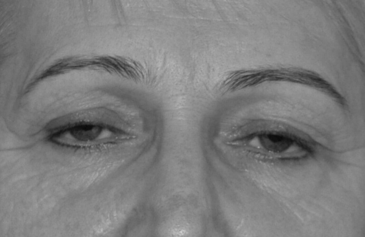
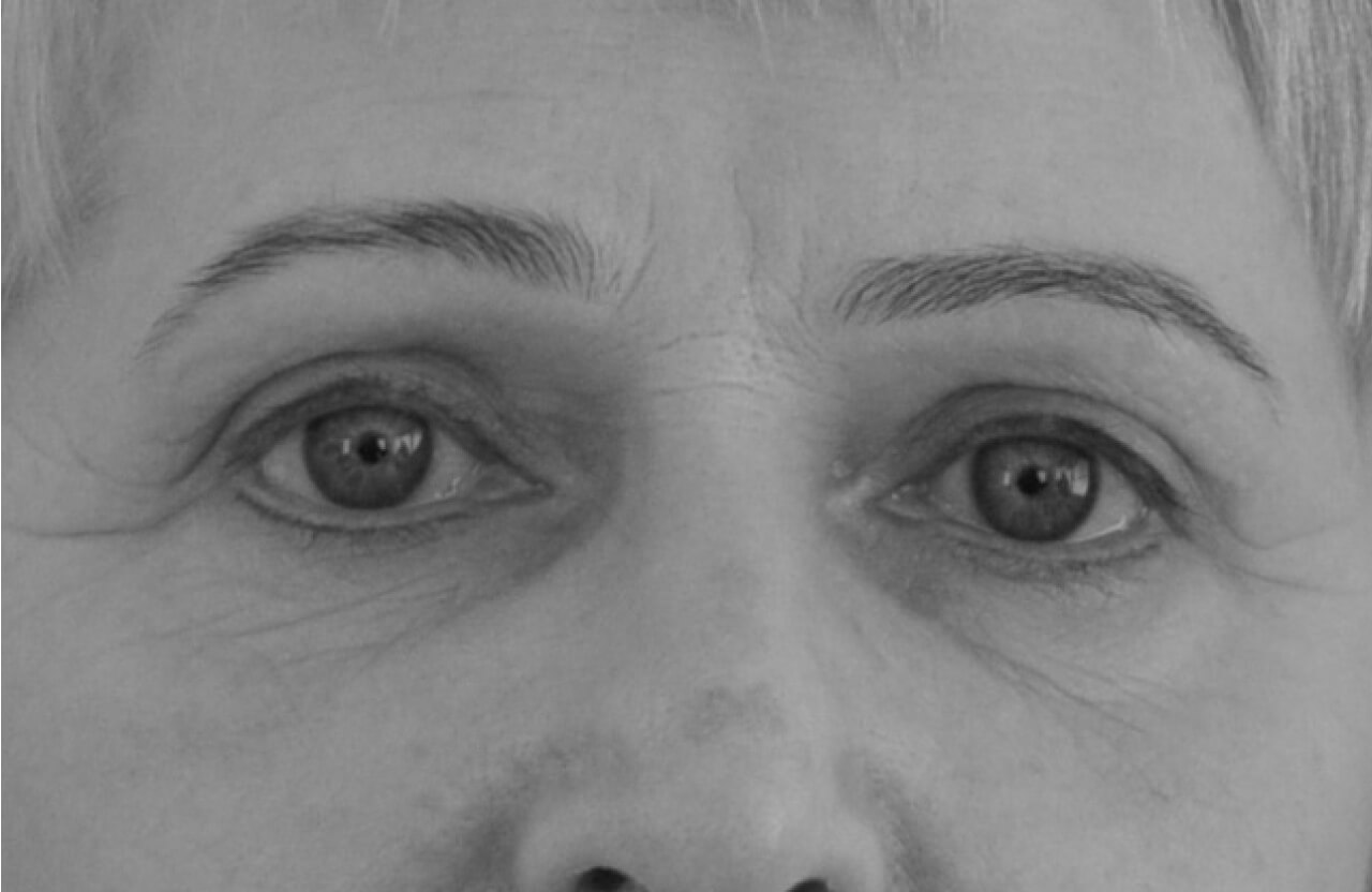
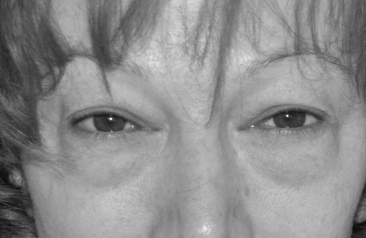
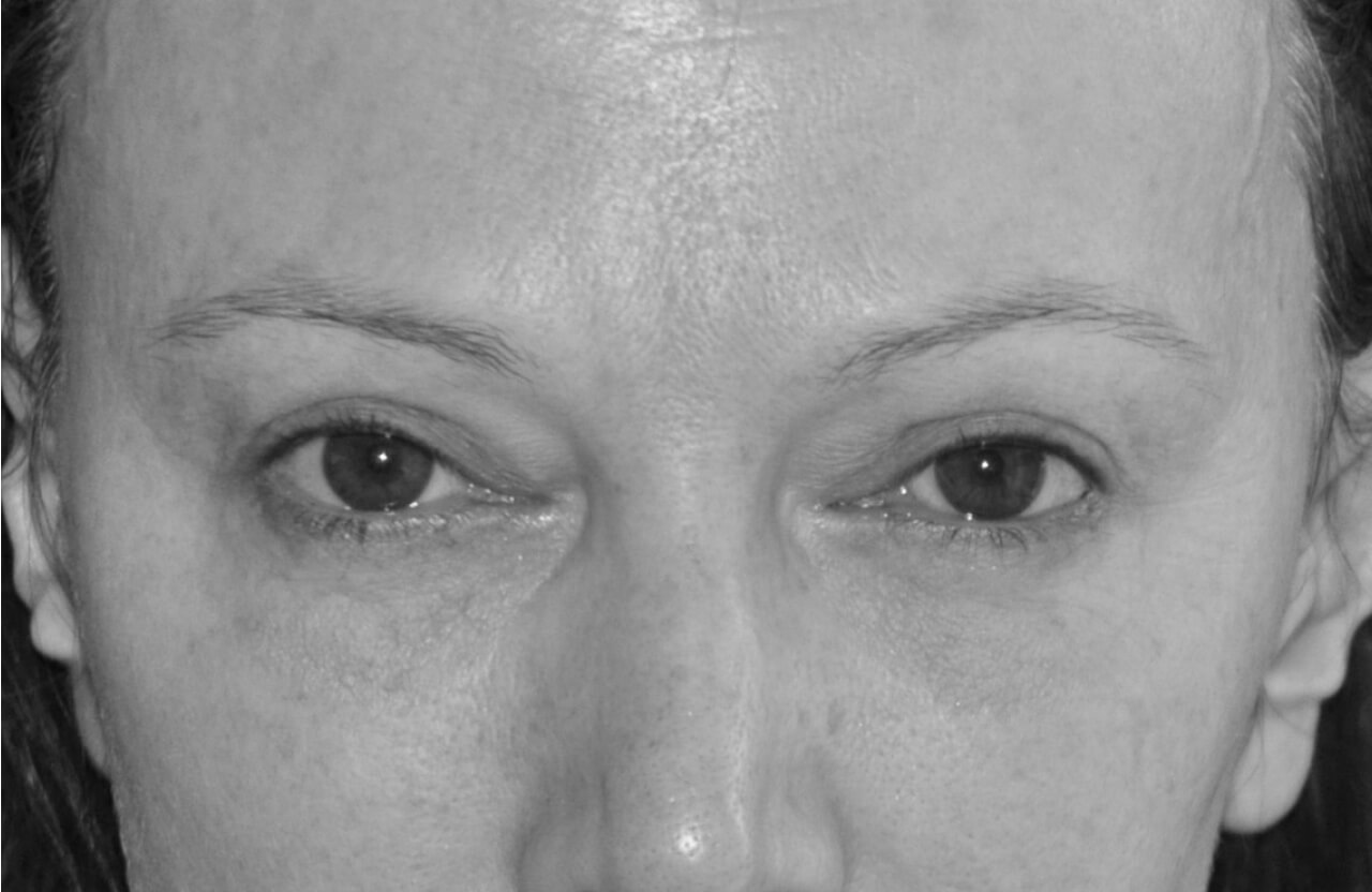
Photo gallery
Detailed information
The human face consists of so-called aesthetic units, such as: forehead, nose, cheeks, eyelids – upper and lower, lips – upper and lower, and chin. Each of these units is perceived against the background of others, and all must fit together. The periorbital area, from a practical point of view, is divided into five zones. Zone I is the upper eyelid, Zone II is the lower eyelid, Zone III is the area of the medial canthus, Zone IV is the area of the lateral canthus, and Zone V is everything that surrounds the eyelids, i.e.: eyebrows, forehead, nose, cheeks. The upper and lower eyelids have a layered structure. The skin of the eyelids is the most superficial layer – it is the thinnest skin in humans. Beneath it is the orbicularis oculi muscle, below that is the tarsus (cartilage that gives the eyelids rigidity), and beneath the tarsus is the conjunctiva, which is the deepest part of the eyelid adhering to the eyeball. The muscle called the levator palpebrae superioris attaches to the upper edge of the tarsus of the upper eyelid. An equivalent of the upper eyelid levator, called the capsulo-palpebral fascia, attaches to the lower edge of the tarsus of the lower eyelid. The orbicularis oculi muscle closes the palpebral fissure, while the levator palpebrae superioris muscle raises the upper eyelid and opens the palpebral fissure. The lower eyelid drops downwards due to gravity and the elasticity of the capsulo-palpebral fascia. A thorough examination and assessment of the anatomical state and function of the eyelids is the basis for success in eyelid plastic surgery.
The upper and lower eyelids are structures that protect the eyeball from injury and drying. The surface of the eyeball should be constantly moist. Under physiological conditions, the surface of the eyeball is covered with a thin layer of tears. This layer is called the „tear film”. Lacrimal glands produce tears, and the movement of the eyelids, called blinking, distributes them evenly over the surface of the eye. We blink 20 times every minute. 1200 times an hour, and about 20,000 times a day. The quantity and quality of tears produced, in addition to eyelid function, are of great importance in protecting the eyeball from drying out. Before eyelid surgery, a detailed medical history regarding past or present eye diseases should be collected from the patient. Patients with a positive history for so-called „dry eye” are at risk of exacerbation of this pathology after surgery. Some doctors routinely refer potential candidates for eyelid surgery for an ophthalmological examination aimed at assessing the quantity and quality of tears.
The upper eyelid in a young person has a small excess of skin. When the eye is open, this excess forms a fold whose edge does not reach the ciliary margin of the eyelid. A strip of eyelid skin above the ciliary margin is visible even with open eyes. With age, the skin slackens, and with open eyes, the skin fold hangs heavily, reaching all the way to the ciliary margin. It sometimes happens that an excessive skin fold of the upper eyelid restricts the field of vision, which causes a reflex in the form of subconscious eyebrow lifting. Raised eyebrows pull the eyelid skin and expose the field of vision. This manifests as constant frowning, which significantly intensifies the formation of wrinkles in this area. True ptosis is not a result of excess skin on the eyelid, but rather insufficient function of the levator palpebrae superioris muscle. The ciliary margin of the upper eyelid then covers part of the pupil when looking straight ahead and restricts the field of vision. Upper eyelid ptosis is often unnoticed or ignored by doctors. A typical aesthetic upper eyelid surgery can improve the field of vision whose restriction results from excess skin, but it will not improve the field of vision resulting from true ptosis. Correction of true ptosis requires surgery on the levator palpebrae superioris muscle. In most cases, shortening this muscle is sufficient to correct the ptosis. Modern upper eyelid surgery involves removing excess skin and orbicularis oculi muscle, removing excess fat in the area of the medial canthus (medial fat herniation) and fat called ROOF (retro orbicularis oculi fat), which is located in the area of the lateral eyebrow arch. If true ptosis is found, this pathology should be corrected. During aesthetic upper eyelid surgery, a fragment of the corrugator supercilii muscle can also be removed to get rid of the so-called „glabellar frown lines”. The procedure can be performed under local anesthesia. The postoperative scar is located in the crease of the upper eyelid, and its lateral part is hidden in one of the wrinkles in the area of the lateral canthus. A single intradermal suture is removed five days after the procedure. For about 7–14 days, the eyelids may be swollen and bruised. For the first few days after the procedure, a calm lifestyle should be maintained, avoiding significant physical exertion and excessive eye strain. It is recommended not to watch television for too long, not to work on a computer, or read books for too long. Sleeping with the head slightly elevated and cold compresses of chamomile essence on the eyelids reduce swelling and help shorten the recovery period. Eye drops used twice a day for 5 days alleviate the postoperative course. Patients do not report pain after upper eyelid surgery, although they are always provided with painkillers, which provides a greater sense of security.
The lower eyelid in a young, healthy person is concave, its vertical (sagittal) dimension is small (the distance from the ciliary margin to the cheek is small), and the transition between the eyelid and the cheek is smooth – the concavity of the eyelid gently transitions into the convexity of the cheek. The skin on a young eyelid is taut. With age, the lower eyelid becomes convex, lengthens in its vertical dimension, and the boundary between the eyelid and the cheek becomes distinct (naso-palpebral groove, also known as „tear trough”, palpebral-malar groove, and so-called „bags under the eyes” – malar bags appear). The eyelid skin becomes increasingly wrinkled. All eyelid structures slacken and become less elastic. The tissues of severely aged lower eyelids hang heavily in the form of festoons, and coexisting senile ectropion can often be observed. Traditional lower eyelid surgery, consisting only of skin removal, is unable to affect most features of the aging lower eyelid and is additionally associated with a fairly high risk of complications. The removal of so-called „fat herniations” from the lower eyelids can correct the convexity of the eyelids and change it to a more aesthetically favorable concavity. Modern lower eyelid surgery corrects all signs of aging and involves repositioning the fat that creates the eyelid convexity towards the tear trough and increasing eyelid tension (cantopexy). The removal of a small excess of skin and fat is only a supplement to the surgery. After surgery, the eyelid is concave, shorter in its vertical dimension, the transition between the eyelid and the cheek is smooth, and the skin is smoothed. The following anatomical features of the lower eyelids pose the greatest risk of postoperative complications:
- negative tilt – this means that in an en face projection, the lateral canthus is lower than the medial canthus;
- negative vector – this means that in a profile projection, the plane of the eyeball is more anterior than the plane of the eyelid and cheek itself;
- eyelid laxity is revealed by three symptoms:
– we are able to pull the ciliary margin of the eyelid more than 8 mm away from the surface of the eyeball;
– after pulling the eyelid away from the eyeball, it slowly returns to its natural position;
– in a ¾ projection (semi-profile), the distance between the lateral canthus and the orbital rim is greater than 1 cm.
These three characteristics (negative tilt, negative vector, and eyelid laxity) are an indication for canthopexy, which means strengthening the eyelid in the horizontal dimension by attaching its lateral margin to the bony orbital rim. If such a procedure is not performed, there is a high probability of a severe complication in the form of eyelid ectropion after surgery.
Modern lower eyelid surgery is performed under local anesthesia, but with the assistance of an anesthesiologist who administers sedatives and, if necessary, applies short-term intravenous anesthesia. The postoperative scar is located just below the ciliary margin of the lower eyelid, and its lateral section is hidden in a crease near the lateral canthus. The wound edges are approximated with single, very thin sutures, which are removed after just five days. After the procedure, plasters are applied to the eyelid to reduce swelling and bruising and to provide support for the eyelid in the early postoperative period. Post-procedure recommendations are the same as after upper eyelid surgery. After the swelling and bruising subside, you can return to your daily activities, and the condition of your eyelids will not reveal the recently performed procedure. Your eyes will look younger and rested. However, the final result of the operation should be awaited for about 3 months. Scars are then very minimally visible, and the eyelids achieve their final appearance.
In the case of very advanced changes in the lower eyelid area (pronounced „bags under the eyes”, eyelid tissues forming garlands), the only effective solution may be the so-called „mid-face lift”. This involves lifting the malar fat pad and suspending it to the bony orbital rim. Lateral canthopexy should always be performed simultaneously. The operation should be performed by an experienced plastic surgeon specializing in eyelid surgery. The procedure, even when performed by the best specialist, carries a risk of complications.
Eyelids are perceived against the backdrop of surrounding aesthetic units, and therefore, correction of the cheek, eyebrows, or nose can favorably influence the appearance of the eyelids. As a result of aging, there is a decrease in the amount of fat tissue in the cheek area, the boundary between the cheek and the eyelid becomes distinct, and there is a lack of a smooth transition between the aesthetic units of the face. Hyaluronic acid preparations or autologous fat grafting are very useful in giving contour and volume to the cheek. By using fillers, one can correct the so-called „tear trough”, mask an overly distinct transition between the cheek and the eyelid, and give the cheeks a rounded shape. These are minimally invasive procedures, without creating permanent external scars, and have a transient effect, meaning the doctor does nothing irreversible. Wrinkles around the lateral canthus („crow's feet”), wrinkles between the eyebrows („frown lines”), and horizontal forehead wrinkles can be softened using botulinum toxin. Skillful injection of the preparation also allows for lifting the lateral parts of the eyebrows upwards, which causes an apparent wider opening of the eye and reduces the amount of skin on the upper eyelid. Minimally invasive procedures using botulinum toxin, hyaluronic acid, and autologous fat are a beneficial complement to surgical eyelid procedures.
Many people focus on an aging neck and lower face, not noticing the changes occurring around the orbits. These changes can be corrected with lower risk and less financial outlay than in the case of a facelift. Moreover, in the case of a facelift focused on the neck and lower face without simultaneous correction of the eyelids and eyebrows, the result of such an operation will further accentuate unfavorable changes around the eyes. On the other hand, aesthetic surgery in the orbital area does not accentuate unfavorable changes resulting from aging in other areas of the face. In other words, one can perform only eyelid surgery without a facelift, but a facelift should not be performed without eyelid surgery.
From an aesthetic point of view, the eyes are probably the most important anatomical unit of the face. When the eyes look fresh and young, the entire face and its aura are fresh, healthy, and full of life. When the eyes look tired, old, and sad, the entire face appears tired, old, and unhealthy. Fortunately, the eye area offers great opportunities for aesthetic improvement. Unfortunately, this same area is where severe complications can occur. There is a dangerous opinion among doctors that blepharoplasty is a quick, simple, and relatively profitable surgical procedure. This is not true. Plastic surgery of the eyelids requires extensive knowledge, skill, and experience. Ensuring patient safety requires dedicating a significant amount of time to gathering medical history and examining the eyelids anatomically and functionally. It is necessary to plan an operation that meets the patient's expectations. The operation itself, lasting about 60 minutes, is just the tip of the iceberg of all pre- and post-procedure activities that contribute to safety.